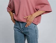
Artinya Hip
Cách tập hip thrust đúng cách:
Một bài tập chân hiệu quả có thể tập cho người mới trước khi tập Squats
Squats là một bài tập giúp tăng cường sức mạnh toàn thân và xây dựng cơ bắp chân hiệu quả. Tuy nhiên, đối với một số người còn hạn chế về mặt thể lực và kỹ thuật tập thì gần như là gặp khó khăn trong khi tập squats.
Để khắc phục điều này, bạn có thể tập hip thrust để thay thế. Bài tập hip thrust cũng cho phép bạn tác động vào nhóm cơ bắp chân khi luyện tập.
Tập hip thrust có tác dụng gì?
Lợi ích nổi bật của bài tập hip thrust là phát triển cơ đùi sau và cơ mông. Bên cạnh đó, bài tập này còn có nhiều tác dụng khác, bao gồm:
Cách tập hip thrust với ghế
Để tập hip thrust với ghế, bạn cần chuẩn bị 1 chiếc ghế bench dài trong phòng gym (flat bench) hoặc 1 chiếc bục chắc chắn và cao khoảng 40 – 60cm.
kb.1 pinggul.2 pangkal paha.-ks. Sl. : modern.-kseru.H.. h. hurrah Hip. hip. hura! Hidup! Horas! -hipped ks. Sl, : keranjingan.
Anatomical region between the torso and the legs, holding the buttocks and genital region
This article is about the anatomical description of the hip. For the cultural description of buttocks, see
. For other uses, see
In vertebrate anatomy, the hip, or coxa[1] (pl.: coxae) in medical terminology, refers to either an anatomical region or a joint on the outer (lateral) side of the pelvis.
The hip region is located lateral and anterior to the gluteal region, inferior to the iliac crest, and lateral to the obturator foramen, with muscle tendons and soft tissues overlying the greater trochanter of the femur.[2] In adults, the three pelvic bones (ilium, ischium and pubis) have fused into one hip bone, which forms the superomedial/deep wall of the hip region.
The hip joint, scientifically referred to as the acetabulofemoral joint (art. coxae), is the ball-and-socket joint between the pelvic acetabulum and the femoral head. Its primary function is to support the weight of the torso in both static (e.g. standing) and dynamic (e.g. walking or running) postures. The hip joints have very important roles in retaining balance, and for maintaining the pelvic inclination angle.
Pain of the hip may be the result of numerous causes, including nervous, osteoarthritic, infectious, traumatic, and genetic.
The hip joint, also known as a ball and socket joint, is formed by the acetabulum of the pelvis and the femoral head, which is the top portion of the thigh bone (femur). It allows for a wide range of movement and stability in the lower body.[3]
The proximal femur is largely covered by muscles and, as a consequence, the greater trochanter is often the only palpable bony structure in the hip region.[4]
The hip joint or coxofemoral joint[5][6] is a ball and socket synovial joint formed by the articulation of the rounded head of the femur and the cup-like acetabulum of the pelvis.[7] The socket of the acetabulum is pointing downwards and anterolaterally. The socket is also turned such that the outer edge of its roof is more lateral than outer edge of the floor.[7] It forms the primary connection between the bones of the lower limb and the axial skeleton of the trunk and pelvis. Both joint surfaces are covered with a strong but lubricated layer called articular hyaline cartilage.
The cuplike acetabulum forms at the union of three pelvic bones — the ilium, pubis, and ischium.[8] The Y-shaped growth plate that separates them, the triradiate cartilage, is fused definitively at ages 14–16.[9] It is a special type of spheroidal or ball and socket joint where the roughly spherical femoral head is largely contained within the acetabulum and has an average radius of curvature of 2.5 cm.[10] The acetabulum grasps almost half the femoral ball, a grip deepened by a ring-shaped fibrocartilaginous lip, the acetabular labrum, which extends the joint beyond the equator.[8] The centre of the acetabulum (fovea) does not articulate to anything. Instead, it is lined with fat pad and attached to ligamentum teres. The acetabular labrum is horse-shoe shaped. Its inferior notch is bridged by transverse acetabular ligament.[7] The joint space between the femoral head and the superior acetabulum is normally between 2 and 7 mm.[11]
The head of the femur is attached to the shaft by a thin neck region that is often prone to fracture in the elderly, which is mainly due to the degenerative effects of osteoporosis.
Transverse and sagittal angles of acetabular inlet plane.
The acetabulum is oriented inferiorly, laterally and anteriorly, while the femoral neck is directed superiorly, medially, and slightly anteriorly.
Acetabular angle (or Sharp's angle)[12] is the angle between the horizontal line passing through the inferior aspects of triradiate cartilages (Hilgenreiner's line) and another line passing through the inferior angle of triradiate cartilage to superior acetabular rim. The angle measures 35 degrees at birth, 25 degrees at one year of age, and less than 10 degrees by 15 years of age.[13] In adults the angle can vary from 33 to 38 degrees.[14]
The sagittal angle of the acetabular inlet is an angle between a line passing from the anterior to the posterior acetabular rim and the sagittal plane. It measures 7° at birth and increases to 17° in adults.[13]
Wiberg's centre-edge angle (CE angle) is an angle between a vertical line and a line from the centre of the femoral head to the most lateral part of the acetabulum,[15] as seen on an anteroposterior radiograph.[16]
The vertical-centre-anterior margin angle (VCA) is an angle formed from a vertical line (V) and a line from the centre of the femoral head (C) and the anterior (A) edge of the dense shadow of the subchondral bone slightly posterior to the anterior edge of the acetabulum, with the radiograph being taken from the false angle, that is, a lateral view rotated 25 degrees towards becoming frontal.[16]
The articular cartilage angle (AC angle, also called acetabular index[17] or Hilgenreiner angle) is an angle formed parallel to the weight bearing dome, that is, the acetabular sourcil or "roof",[18] and the horizontal plane,[15] or a line connecting the corner of the triangular cartilage and the lateral acetabular rim.[19] In normal hips in children aged between 11 and 24 months, it has been estimated to be on average 20°, ranging between 18° and 25°.[20] It becomes progressively lower with age.[21] Suggested cutoff values to classify the angle as abnormally increased include:
The angle between the longitudinal axes of the femoral neck and shaft, called the caput-collum-diaphyseal angle or CCD angle, normally measures approximately 150° in newborn and 126° in adults (coxa norma).[23][dubious – discuss]
An abnormally small angle is known as coxa vara and an abnormally large angle as coxa valga. Because changes in shape of the femur naturally affects the knee, coxa valga is often combined with genu varum (bow-leggedness), while coxa vara leads to genu valgum (knock-knees).[24]
Changes in the CCD angle is the result of changes in the stress patterns applied to the hip joint. Such changes, caused for example by a dislocation, change the trabecular patterns inside the bones. Two continuous trabecular systems emerging on the auricular surface of the sacroiliac joint meander and criss-cross each other down through the hip bone, the femoral head, neck, and shaft.
On the lateral side of the hip joint the fascia lata is strengthened to form the iliotibial tract which functions as a tension band and reduces the bending loads on the proximal part of the femur.[23]
Proximally, capsule of the hip joint is attached to the edge of the acetabulum, acetabular labrum, and transverse acetabular ligament. Distally, it is attached to the trochanters of the femur and intertrochanteric line anteriorly. Posteriorly, it is attached to a junction between medial two-thirds and lateral one-third of the femoral neck,[7] one finger breadth away from the intertrochanteric crest.[24] From its attachment at the femoral neck, the fibres of the capsule reflected backwards towards the acetabulum, carrying retinacula vessels supplying the femoral head.[7] The part of femoral neck outside the capsule is shorter in front than posteriorly.[24]
The strong but loose fibrous capsule of the hip joint permits the hip joint to have the second largest range of movement (second only to the shoulder) and yet support the weight of the body, arms and head.
The capsule has two sets of fibers: longitudinal and circular.
Extracapsular ligaments. Anterior (left) and posterior (right) aspects of right hip.
Intracapsular ligament. Left hip joint from within pelvis with the acetabular floor removed (left); right hip joint with capsule removed, anterior aspect (right).
The hip joint is reinforced by four ligaments, of which three are extracapsular and one intracapsular.
The extracapsular ligaments are the iliofemoral, ischiofemoral, and pubofemoral ligaments attached to the bones of the pelvis (the ilium, ischium, and pubis respectively). All three strengthen the capsule and prevent an excessive range of movement in the joint. Of these, the Y-shaped and twisted iliofemoral ligament is the strongest ligament in the human body. It has a tensile strength of 350 kg.[24] Iliofemoral ligament is a thickening of the anterior capsule extending from anterior inferior iliac spine to intertrochanteric line.[7] Ischiofemoral ligament is the thickening of posterior capsule of the hip and pubofemoral ligament is the thickening of the inferior capsule.[7] In the upright position, iliofemoral ligament prevents the trunk from falling backward without the need for muscular activity, thus preventing excessive hyperextension. In the sitting position, it becomes relaxed, thus permitting the pelvis to tilt backward into its sitting position. Ischiofemoral prevents excessive extension and the pubofemoral ligament prevents excess abduction and extension.[26]
The zona orbicularis, which lies like a collar around the most narrow part of the femoral neck, is covered by the other ligaments which partly radiate into it. The zona orbicularis acts like a buttonhole on the femoral head and assists in maintaining the contact in the joint.[24] All three ligaments become taut when the joint is extended - this stabilises the joint, and reduces the energy demand of muscles when standing.[27]
The intracapsular ligament, the ligamentum teres, is attached to a depression in the acetabulum (the acetabular notch) and a depression on the femoral head (the fovea of the head). It is only stretched when the hip is dislocated, and may then prevent further displacement.[24] It is not that important as a ligament but can often be vitally important as a conduit of a small artery to the head of the femur, that is, the foveal artery.[28] This artery is not present in everyone but can become the only blood supply to the bone in the head of the femur when the neck of the femur is fractured or disrupted by injury in childhood.[29]
The hip joint is supplied with blood from the medial circumflex femoral and lateral circumflex femoral arteries, which are both usually branches of the deep artery of the thigh (profunda femoris), but there are numerous variations and one or both may also arise directly from the femoral artery. There is also a small contribution from the foveal artery, a small vessel in the ligament of the head of the femur which is a branch of the posterior division of the obturator artery, which becomes important to avoid avascular necrosis of the head of the femur when the blood supply from the medial and lateral circumflex arteries are disrupted (e.g. through fracture of the neck of the femur along their course).[29]
The hip has two anatomically important anastomoses, the cruciate and the trochanteric anastomoses, the latter of which provides most of the blood to the head of the femur. These anastomoses exist between the femoral artery or profunda femoris and the gluteal vessels.[30]
Cách tập hip thrust với máy (machine)
Thay vì bạn phải chuẩn bị tạ đơn hoặc tạ đòn để tập thì có một số phòng tập đã trang bị sẵn máy chuyên dụng để tập hip thrust. Bài tập này còn được gọi là ‘Hip thrust machine quick tutorial’.
Cách thực hiện tương tự như những cách tập của những biến thể khác.
Cách tập hip thrust với tạ đòn
Sau khi bạn đã nắm được một vài kỹ thuật căn bản thì bài tập hip thrust với tạ đòn (barbell hip thrust) cũng tương đối dễ thực hiện. Khi sử dụng thanh tạ đòn, bạn lưu ý chọn mức tạ vừa đủ, vì khi thực hiện động tác gập hông thanh tạ đòn sẽ di chuyển và khiến bạn mất thăng bằng.
Thay vì dang rộng hai cánh tay sang hai bên thì đối với bài tập này bạn sẽ dùng hai tay để giữ thanh tạ sao cho không trượt ra khỏi cơ thể.
Lưu ý khi tập hip thrust
Theo thông tin từ Tạp chí cung cấp thông tin về ăn uống và tập luyện – LiveStrong, các chuyên gia thể dục thể thao đưa ra một vài lưu ý để chúng ta có thể tập hip thrust hiệu quả hơn.
Tóm lại, hip thrust là một bài tập cho phần thân dưới, giúp gia tăng sức mạnh và giúp phát triển vòng 3 hiệu quả. Bên cạnh việc tìm hiểu về bài tập, bạn cần nhớ rằng, để hoàn thành bài tập hiệu quả, bạn nên bắt đầu với mức tạ vừa phải, tập trung vào kỹ thuật tập thay vì cố gắng lên tạ quá mức.
[embed-health-tool-bmi]
Clinical significance
A hip fracture is a break that occurs in the upper part of the femur.[33] Symptoms may include pain around the hip particularly with movement and shortening of the leg.[33] The hip joint can be replaced by a prosthesis in a hip replacement operation due to fractures or illnesses such as osteoarthritis. Hip pain can have multiple sources and can also be associated with lower back pain.
At the 2022 Consumer Electronics Show, a company named Safeware announced an airbag belt that is designed to prevent hip fractures among such uses as the elderly and hospital patients.[34]
Abnormal orientation of the acetabular socket as seen in hip dysplasia can lead to hip subluxation (partial dislocation), degeneration of the acetabular labrum. Excessive coverage of femoral head by the acetabulum can lead to pincer-type femoro-acetabular impingement (FAI).[7]
Cách tập hip thrust với tạ đơn
Tập hip thrust với tạ đơn (dumbbell hip thrust) cũng tương tự như bài tập hip thrust với tạ đòn, chỉ khác nhau ở chỗ thay thanh tạ đơn dài bằng quả tạ đơn. Bài tập này phù hợp cho những bạn mới bắt đầu tập, vì tạ đơn tương đối dễ kiểm soát và dễ giữ thăng bằng hơn so với tập tạ đòn.
Khi thực hiện động tác, lưu ý phần đầu của tạ đơn phải được đặt gần vị trí đặt hông, đồng thời dùng 2 tay cố định hai đầu của tạ.


